


Introduction
Obstructive sleep apnea (OSA) is a common affliction caused by intermittent obstruction of the upper respiratory tract. Despite the effort to breathe, there are repeated cessations of normal breathing. These cessations are termed as apneas. The resultant apnea causes sleep fragmentation, which in turn increases sympathetic activity, decreases insulin sensitivity and glucose uptake, and stimulates hepatic gluconeogenesis that ultimately leads to type 2 diabetes [1]. Type 2 diabetes is a major chronic disease with high morbidity, mortality, and economic burden [2]. Obesity, one of the prime reason for the alarming rise in the prevalence of type 2 diabetes [3,4]. However, recent evidence based, clinical based, and laboratory based studies suggested that diabetes and OSA are independently associated with degree of adiposity [4]. A few laboratory studies in healthy young subjects have found that, under well-controlled conditions, restricting sleep duration had an adverse impact on glucose tolerance [5,6]. These studies suggest that treatment of OSA condition will ultimately improve glucose metabolism and eventually will lead to improved glucose control, however the effect of OSA treatment on glucose control has been variable.
Review of the literature found that most studies exploring the effect of continuous positive airway pressure (CPAP) on insulin sensitivity show a positive effect [3], however literature lack evidences on the effect of mandibular advancement device (MAD) on insulin resistance (IR). Hence the present study was planned to evaluate the effect of mandibular advancement splint (MAS) on IR in patients with OSA.
Methods
The present study was conducted at Department of Prosthodontics, Dental College Azamgarh, from June 2015 to July 2017. Ethical approval was obtained from Committee for Ethics in Human Research Azamgarh. All the patients were informed about the nature of the study and written informed consent was taken from all the patients.
The inclusion criteria were as follows: 1) OSA patients with type 2 diabetes mellitus with stable diabetic control under oral hypoglycaemic medication (sulfonylurea or alpha-glucosidase inhibitor), 2) HbA1c value >5.8% since in India the cutoff value of HbA1c for the patient to have diabetes is >5.8% [7], 3) age range 35 to 50 years, and 4) non obese (body mass index 18–25 kg/m2). The exclusion criteria were as follows: 1) patients on insulin therapy, 2) apnea-hypopnea index (AHI) >30 since oral appliances are more effective in mild and moderate OSA, 3) craniofacial abnormalities (cleft lip and palate, retruded mandible) since these might act as confounding factors by further accentuating the condition of OSA, and 4) mobile tooth and temporomandibular joint disorders including pain, significant joint crepitation, restricted mouth opening, and masticatory muscle tenderness since they effect the normal functioning of MAS.
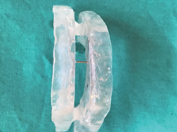
The primary screening yielded a cohort 92 patients who were assessed for excessive daytime sleepiness and sleep disordered breathing by the Epworth Sleepiness Scale and Berlin Questionnaire, respectively. 86 symptomatic patients were advised to undergo all night polysomnography. Based on AHI, patients were divided into mild (n=28), moderate (n=44), and severe (n=14) grade of OSA. Severe grade OSA patients were excluded from the study. For the remaining patients (n=72), a MAS was fabricated and was fixed at 70% of the maximum mandibular protrusion recorded, to avoid any possible anterior impingement of the glenoid fossae by the condyles (Fig. 1) [8]. All patients were recalled every week till one month. Four patients did not report. Patients that were comfortable with MAS after one month (n=68) were assessed for AHI, mean oxygen (O2) saturation and IR at baseline, 6 months, and 1 year after wearing MAS. Thus the sample size constituted twenty eight mild OSA patients and forty moderate OSA patients. A self reported adherence to the oral appliance therapy was assessed every week via telephone. No patients were lost in follow-up. Demographic data is presented in Table 1.
AHI and mean O2 saturation
A full night polysomnography was performed in the sleep laboratory by sleep technician using S-7000 computerized polysomnography machine (Cogent Technologies, Woodbridge, U.K.) that included electroencephalograms, electrooculogram, chin and leg electromyogram, nasal airflow (nasal pressure cannula), O2 saturation (pulse oximetry), movements of thorax and abdomen, electrocardiogram, and body position. Somnologica Studio software (Embala corporation, Thornton, CO, USA) was used to calculate AHI in accordance with American Academy of Sleep Medicine manual for the scoring of sleep and associated events, version 2.0 [9]. AHI was determined by the frequency of these events per hour during sleep time based on the results of the overnight polysomnography [8].
IR
Venous blood was sampled in the morning after an overnight fast for the measurement of the values of plasma glucose, serum insulin, and HbA1c. Plasma glucose was measured by glucose oxidase method, serum insulin was measured with radioimmunoassay, and HbA1c values were measured by high performance liquid chromatography. Homeostasis model assessment (HOMA) was used to estimate IR [10]. HOMA-IR was calculated with the formula: fasting plasma glucose (mmol/L) times fasting serum insulin (mU/L) divided by 22.5. Low HOMA-IR values indicate high insulin sensitivity, whereas high HOMA-IR values indicate low insulin sensitivity. The normal range of HOMA-IR value is 0.5–1.4 in healthy individual. HOMA-IR value above 2.9 indicates significant IR [11,12].
Data was analyzed using Statistical Package for Social Sciences Version 24.0 (IBM Corp., Armonk, NY, USA). Analysis of variance statistical analysis was done with paired t-test to assess the significance of mean difference at different time points within each OSA severity sub-group for all three parameters (AHI, mean O2 saturation, and HOMA-IR value). Pearson correlation analysis was used to correlate between AHI and other parameters. A p value less than 0.05 indicated statistically significant difference.
Results
The baseline, 6 months, and 1 year AHI scores, mean O2 saturation, and HOMA-IR values are given in Table 2. A statistically significant reduction in mean AHI was observed after 6 months and 1 year of wearing oral appliance (p=0.051 and p<0.01, respectively) in both mild and moderate OSA patients.
No statistically significant improvement in mean O2 saturation was observed after 6 months of MAS use (p=0.061 for mild OSA patients and p=0.072 for moderate OSA patients); however long term use (1 year) of MAS showed a statistically significant improvement in mean O2 saturation in both mild as well as moderate OSA patients (p=0.05 and p=0.002, respectively).
For mild OSA patients a statistically significant reduction in HOMA-IR value was observed at 6 months and 1 year (p=0.001 and p=0.002, respectively); however for moderate OSA patients no significant improvement was observed following MAS use (p=0.306 at 6 months and p=0.172 at 1 year).
On seeking the correlation between AHI, mean O2 saturation, and HOMA-IR value at 6 months, only HOMA-IR value showed a significant positive correlation with AHI (r=0.547; p=0.03) in mild OSA patients (Table 3). No significant correlation was observed for moderate OSA patients (Table 3). At 1 year, HOMA-IR value showed a moderate positive correlation with AHI in mild as well as moderate OSA patients (r=0.522, p=0.04; r=0.445, p=0.03, respectively) (Table 4).
Discussion
The present pilot study was aimed at evaluating the effect of oral appliance on IR on a cohort of OSA patients with type 2 diabetes and who were not on insulin therapy and had stable diabetic regime. We hypothesized that MAS will not have any effect on insulin sensitivity in OSA patients.
The results of the study indicated that 6 months of MAS use significantly decreases IR in mild OSA patients. No significant improvement in insulin sensitivity was observed in moderate OSA patients with 6 months use of oral appliance; however long term use showed significant improvement in IR. This might be due to the effectiveness of oral appliance in treating mild OSA patients as compared to moderate OSA patients [13-15]. Although there was improvement in HOMA-IR value but there was no normalisation [11]. The maximum improvement was observed in mild OSA group that used MAS for 1 year. Bleifeld et al. showed that the longer the duration of CPAP use, the better was the insulin sensitivity [16]. Similar observation was reported by Harsch et al. that treatments that improve OSA will also improve insulin sensitivity in diabetic subjects with OSA [17], although this improvement seems to take somewhat longer than in nondiabetic subjects. Wang and Wei et al. showed that short term use of CPAP could improve the hypoxia of the patients and it could enhance the insulin sensitivity [18,19]. Although CPAP is the gold standard for treating OSA, effectiveness of oral appliance in mild OSA has also been established.
Another finding of the study was reduction in AHI and improvement in mean O2 saturation in mild and moderate OSA patients. The most commonly employed measure of OSA is AHI. Our findings are consistent with Gao et al. who found that after intervention by MAD, AHI decreased significantly [20]. Our findings agree with those of Liu et al. and Ranieri et al. who found statistically significant reduction in AHI score after MAD intervention [21,22]. Studies have observed that a reduction in AHI score was related to an increased velopharynx cross-sectional area after use of MAD.
Thus our hypothesis was rejected as there was the improvement in insulin sensitivity in OSA patients after intervention with MAS. This study provides evidence to inform health care worker about possible use of MAS in OSA with type 2 diabetes mellitus. To our knowledge, this study is the first to objectively evaluate the oral appliance effect on IR. Limitations of the present study include absence of any controls that would have correlated between normal OSA patients and patients with type 2 diabetes mellitus. Another limitation was Somnologica Studio software was used to calculate AHI without being reviewed by a physician. Although there are studies predicting the reliability of automated scoring of polysomnographic data over manual scoring [23-25], further studies are required before using this software alone. Another limitation was the effect of lifestyle factors and obesity, which were not part of the study.
In conclusion, the finding suggested that oral appliance is effective in improving IR in mild OSA patients. The severity of OSA has been associated with IR and subsequent impaired glucose metabolism, however further research in this field is warranted.