


Introduction
Central neurocytoma is rare, neuronal differentiated tumors and comprise about less 1% of intracranial tumors. Central neurocytoma is usually located within the ventricular system and presents symptom as increasing obstructive hydrocephalus. The most common symptom is headache, dizziness, nausea, vomiting, papilledema, and ataxia by increased intracranial pressure [1]. Here we report a rare case of central neurocytoma presenting as symptomatic cataplexy.
Case Report
A 50-year-old man with a history of hypertension and hyperlipidemia visited our clinic complaining of repetitive drop down, which started six months ago. The paralytic attacks were mainly triggered by emotional changes. He has been managing a billiard room for 10 years and had very good billiard skills. When the patient concentrates on a very difficult 3-balls billiards game and solves the problem by hitting the ball correctly, a paralytic attack has occurred followed by a virtuous feeling. He also complained of mild excessive daytime sleepiness from three years ago. Epworth sleepiness scale was 8 and Stanford sleepiness scale 3. There was no preceding history of encephalitis, antipsychotic medication use, or exposure to toxic substances. A neurological examination was normal.
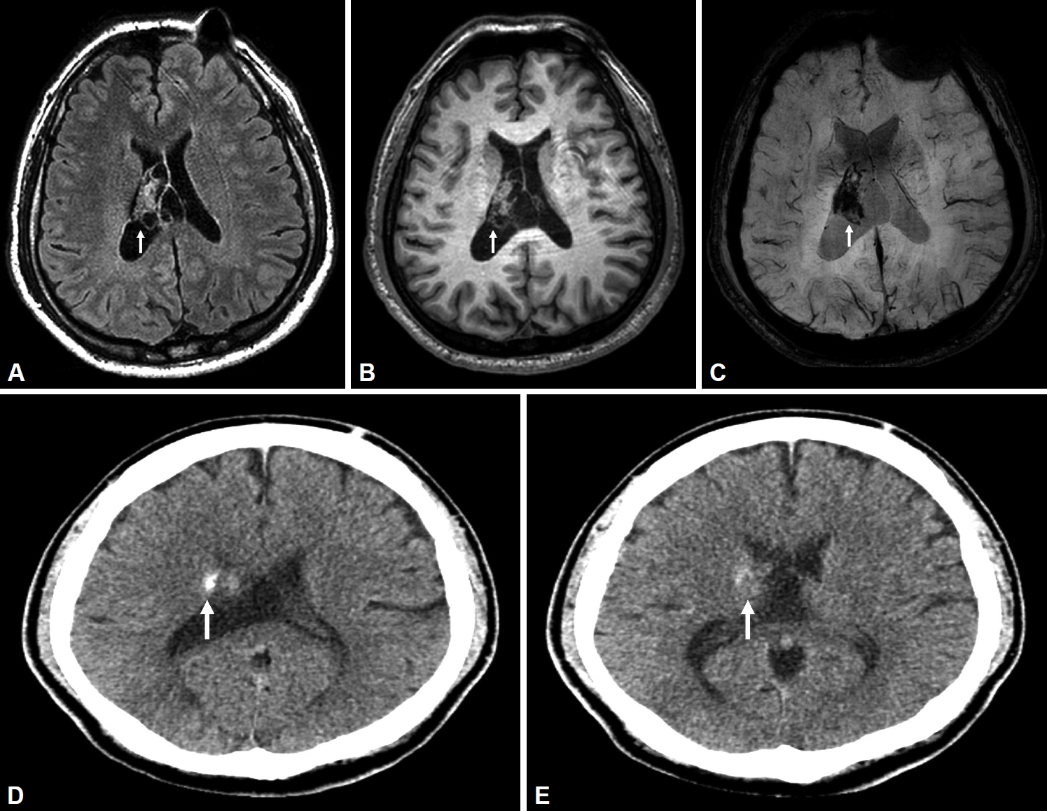
Since the patient’s symptoms occurred in abruptly repeated pattern triggered by emotional changes during wakefulness, cataplexy was suspected, and a laboratory and imaging diagnostic workup was performed. There were no significant laboratory abnormalities including serology and hematological tests, thyroid function tests, rapid plasma reagin, and Treponema pallidum hemagglutination assays. An electroencephalogram was reported as normal. However, brain magnetic resonance imaging demonstrated tumor with calcification occupying right lateral ventricles on fluid-attenuated inversion recovery, T1-weighted imaging, and susceptibility-weighted imaging sequences (Fig. 1A-C). In addition, there was a calcified lesion at the right head of caudate nucleus on brain computed tomography of the patient (Fig. 1D, E). We tried to perform a polysomnography for further evaluation, he refused the examination and we lost to additional follow-up.
Discussion
To the best of our knowledge, a central neurocytoma presenting as symptomatic cataplexy has never been reported. Central neurocytomas are well-differentiated tumors and are typically found in the lateral or third ventricle, attached to the septum pellucidum or ventricular wall at the foramen of Monro [2]. Most patients present with symptoms of increased intracranial pressure due to hydrocephalus such as headache, dizziness, and visual disturbances, but focal neurologic deficits are uncommon.
Cataplexy is characterized as sudden involuntary muscle weakness or paralysis during wakefulness, typically triggered by strong emotions, and is the indispensable symptom of narcolepsy with cataplexy [3]. It has been known that cataplexy occurs almost exclusively in patients with depletion of hypothalamic hypocretin neurons. Although the fascinating link between emotion and cataplexy remains unclear, the hypocretin system has strong reciprocal links with limbic areas such as the dopaminergic ventral tegmental area and the amygdala. These areas primarily involved in reward and emotional processing [4]. Patient with cataplexy has been known to have abnormal activity in reward brain circuit [5].
Alterations of striatal dopaminergic system in narcolepsy with cataplexy also have been reported. In functional brain study, patients with narcolepsy showed increased striatal D2 receptor binding and a positive correlation between this and the frequency of cataplexy and sleep attack [6]. Besides, in cerebral perfusion measured by brain single photon emission computed tomography showed hypoperfusion in bilateral caudate nuclei [7]. In patient with cataplexy, the hypothalamus is the most common region, but there are no reports associated with single brain lesion, except multiple white matter lesion in patient with multiple sclerosis [8]. The limitation of this case is lack of histological confirmation and clinical change due to no surgery. Through this case, we suggest the striatal lesion might play a role in cataplexy.